Sacroiliac Joint – Part 1
Sacroiliac Joint – Part 1
The Sacroiliac Joint (SIJ), of which we have two, is the joint articulation between the sacrum and the two pelvic bones. The SIJs are essential for effectively transferring loads between the lower limbs and the spine. They are highly specialized joints that permit stable (yet flexible) support to the upper body. The sacrum, pelvis and spine, and the connections to the arms, legs and head are functionally interrelated through muscular, fascial and ligamentous interconnections (Vleeming et al, 2012). This means that when you experience pain in the pelvic region we need to consider the pelvis, spine and limbs as an integrated, interdependent and dynamic biological structure, not just focus on the painful spot. However the most likely cause of a specific SIJ injury is “jarring” of the leg if coming off a step or a sudden force going through the limb when standing on one leg.
Many ligamentous, fascial and musculous tissue cross the SIJs but none specifically work on the joints. The main muscles that do have an indirect impact on the SIJ are: gluteus maximus and minimus, psoas, multifidis, pelvic floor muscles and the diaphragm, transversus abdominus, latissimus dorsi, piriformis, biceps femoris of the hamstring group, thoracolumbar fascia, external and internal obliques.
The SIJ is encased in a capsule that has a smooth anterior wall and irregular bands comprising the posterior wall. The capsule has a nerve supply and is surrounded by several strong ligaments which influence its range of motion. In turn, these ligaments are related to the thoracolumbar fascia which is itself derived from fibres of several larger back muscles that surround the joint at a distance.
Current research now supports the existence of very limited ranges of motion of the SIJs in all three of their planes. The average range is 2 ° of movement (Goode et al, 2008; Kinsgard et al, 2017).
Research has shown that increased SIJ laxity is not associated with pregnancy-related pelvic pain and that pregnant women with moderate or severe pelvic pain have the same laxity in the SIJs as pregnant women with no or mild pain. (Damen et al, 2001). Research has also found that there are no provoking or relieving movements or positions that are unique or especially common to SIJ pain therefore other structures, above or below, may be the source of pain. Reliability of palpation testing for SIJ pain has been found to be poor (Robinson et al, 2007) which means palpating the SIJ under the thoracolumbar fascia and the surrounding muscles and ligaments is not useful in telling use that the SIJ is the source of pain.
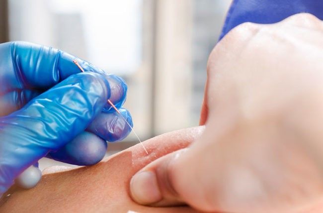
Diagnosis of SIJ pain
The current gold standard for diagnosing SIJ pain is to use fluoroscopically-guided, contrast enhanced intra-articular anaesthetic nerve blocks (Laslett et al, 2005) – which we obviously cannot do in a physiotherapy clinic! From a physiotherapy assessment point of you, the tests that are both clinically effective and backed by best evidence available is a group of tests called Pain Provocation Tests. These consist of six specific tests that put the SIJ under stress in different ways. If three or more of these tests are painful, we can deduce that the SIJ is the source of pain. If there are less than three tests positive or if none of the six tests induce pain, we can confidently say that the SIJ is not the source of pain. We also need to assess the joints and musculature above and below the SIJ to get a complete picture. Treatment options and exercises for SIJ-related pain will be discussed in Part 2.

Aine Tunney MISCP
References
- Damen, L., Buyruk, H.M., Guler-Uysal, F., Lotgering, F.K., Snijders, C.J., Stam H.J. (2001) Pelvic pain during pregnancy is associated with asymmetric laxity of the sacroiliac joints. Acta Obstet Gynecol Scand , 80(11): 1019-24.
- Goode, A., Hegedus, E.J. et al (2008) Three-dimensional movements of the sacroiliac joint: a systematic review of the literature and assessment of clinical utility. The Journal of Manual and Manipulative Therapy , 16(1):25-38.
- Kibsgard, T.J., Rohrl, S.M. et al (2017) Movement of the sacroiliac joint during the Active Straight Leg Raise test in patients with long-lasting severe sacroiliac joint pain. Clinical Biomechanics , 47:40-45.
- Laslett, M., Aprill, C.N. et al (2005) Diagnosis of sacroiliac joint pain: validity of individual provocation tests and composites of tests. Manual Therapy, 10:207-218.
- Robinson, H.S., Brox, J.I. et al (2007) The reliability of selected motion- and pain provocation tests for the sacroiliac joint. Manual Therapy , 12:72–79.
- Vleeming, A., Schuenke, M.D. et al (2012) The sacroiliac joint: an overview of its anatomy, function and potential clinical implications. Journal of Anatomy , 221:537-567.